Keloids: Too Much of a Good Thing!
A keloid scar is an exuberant growth of scar tissue that forms at sites of skin injury. The term keloid, which means "crab-claw", was first used by Alibert in 1806 in reference to the way that the lesions expand from the original scar laterally into the normal adjacent tissue. Keloid scars are differentiated from hypertrophic scars in that hypertrophic scars stay within the boundaries of the cutaneous insult and may spontaneously regress, whereas keloids expand into normal adjacent tissue and do not regress. Keloids and keloid scars occur as a result of a variety of cutaneous insults, including piercings, tattoos, acne, vaccinations, surgery, infection and trauma.
Keloid scars can be distressing to patients psychologically because of their appearance. They are often pruritic and/or painful, and can be physically restricting if located over joints. A recent study found that keloids affect patients' quality-of-life as profoundly as psoriasis does to psoriatics, underscoring how much suffering can accompany this condition.
Epidemiology and Genetics
Keloid scarring can occur at any age but most commonly appear between the ages of 10 and 30 years. The prevalence can be as high as 16% in those of African descent; Hispanic and Asians also have a higher prevalence rate compared with Caucasians. Keloids are most common on the chest, shoulders, back and earlobes, and are least commonly seen on the palms and soles. A slight female predilection has been suggested but this may be due to women piercing their ears more often than men. Keloids can occur within families; most of the studies suggest autosomal-dominant inheritance with variable expression and incomplete penetrance, although one study describes autosomal recessive inheritance.
Given the racial differences and familiar inheritance patterns, there is ongoing research towards discovering the genes that predispose to keloid formation. Although transforming growth factor-β (TGFβ) signaling has been implicated in keloid formation, no polymorphisms have been found within the genes for TGFβ1, TGFβ2, or the TGFβ receptor. A genome-wide association study and linkage analyses of multiple affected families have found several distinct regions of the human genome that are highly correlated with keloid formation. These results highlight the complexity underlying the pathogenesis of this disorder.
Diagnosis
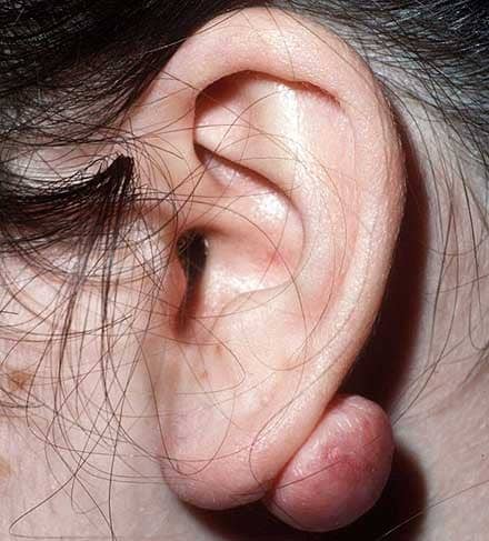
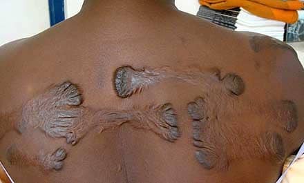
Most keloid scars are round or oval with regular borders; on the ears or neck, they tend to be pedunculated (Figure 1). Those on the chest, back and extremities are usually raised with a flat surface, and tend to have claw-like configurations with irregular borders (Figure 2). Keloids can be soft and doughy or hard and rubbery. Early lesions can be erythematous in color, but later on they often become hyperpigmented, especially in skin of color. The diagnosis of keloids is usually made clinically; however, conditions that mimic keloids include dermatofibrosarcoma protuberans (DFSP), lobomycosis and cutaneous leiomyomas.
Histologically, keloids have abnormally broad, homogeneous, hyalinized collagen bundles arranged in an irregular, disorganized fashion. An increased number of fibroblasts, mast cells and mucin are found in between the collagen bundles. Keloids have less vascularity compared with hypertrophic scars and normal healing skin. The epidermis may be atrophic, and there is often an inflammatory infiltrate around telangiectatic vessels in the upper dermis.
Treatment Options
No single modality is 100% effective for the treatment of keloid scars, and so prevention is better than cure. Non-essential surgeries, piercings and tattoos should be avoided in individuals with a personal or family history of keloid formation, especially at predilection sites. When surgery must be performed, efforts should be made so that closures occur along the relaxed skin tension lines (to reduce skin tension). Although effective, surgical excision alone has recurrence rates ranging from 45-100%, with the recurrent keloid often growing faster and larger than the previous lesion. Combining surgical excision with other treatment modalities (see below) leads to improved outcomes, with recurrence rates decreasing to 8-50%.
Corticosteroids have been a relatively effective first-line treatment modality for keloids: they decrease collagen formation and promote increased collagenase activity. Triamcinolone at a concentration of 10-40mg/mL is injected into the lesion intradermally every 4-6 weeks for several treatments or until the lesion has flattened. Although not completely removed, the keloids often become smaller, softer and less symptomatic. Intralesional corticosteroids are often administered immediately after keloid excision to decrease the chance of recurrence. The main side effects are injection pain, dyspigmentation, and skin/fat atrophy.
Silicone sheets are worn over the surgical scar for 8-24 h a day for several months. Their mechanism of action is thought to be secondary to occlusion and hydration.
Mechanical compression has been used primarily to treat earlobe keloids. After surgery, compression devices are used, which are most effective if worn 24 h/day for several months at a pressure level of 25 mmHg. The mechanism of action is unknown but is believed to be a result of decreased oxygen tension in the wound owing to occlusion of smaller blood vessels.
Radiation therapy is usually used in combination with surgical excision and is usually given within the first 2 weeks post-excision, during the proliferative phase of wound healing. The main side effects are dyspigmentation, dermatitis and telangiectasias. Larger doses of therapy (usually administered as monotherapy) carry an increased risk of future skin cancer development.
Cryotherapy has been used but pain and depigmentation are concerning side effects. Nevertheless, cryotherapy combined with intralesional steroids has shown a response rate as high as 84%.
Newer keloid treatment options include intralesional interferon, which has been shown to reduce collagen and mucin production and increase collagenase activity. This is best performed immediately after surgery, with an injection of 1x10⁶ U/cm of skin surrounding the surgical site. The injections are repeated every 1-2 weeks and can reduce the size of formed keloids by up to 50%.
Smaller keloids can be treated with intralesional 5-fluorouracil (5-FU). The optimal regimen is a mixture of one part triamcinolone (10 mg/mL) to nine parts 5-fluorouracil (50 mg/mL) injected three times weekly.
Various lasers have been used to treat keloids; however, recurrence rates can be greater than 90%.4 Case reports have reported efficacy of methotrexate and colchicine in preventing the recurrence of keloids post-excision; however, larger studies are required to validate these initial findings.
A follow-up period of at least 1 year is recommended, as none of the above therapies come without a significant risk of recurrence; this risk illustrates that more scientific research is required to uncover the molecular mechanisms underlying keloidogenesis.
Conclusions
Keloids are the result of excessive wound healing of the skin. Although surgical excision, intralesional corticosteroids, and radiation therapy remain the mainstay therapeutic options, a number of new treatment modalities exist but need validation before they can be recommended with confidence. To date, there is no keloid treatment that is 100% effective in preventing or treating keloids; however, additional research into the genetic causes of keloid formation is ongoing.