Blurred lines: Erythema Dyschromicum Perstans and Lichen Planus Pigmentosus
The terms lichen planus pigmentosus (LPP), erythema dyschromicum perstans (EDP) and idiopathic eruptive macular pigmentation (IEMP) are often used interchangeably, and if one examines the medical literature, what is referred to as LPP in one publication is termed EDP or IEMP in another. Even pigmentary disorder experts around the world differ in their opinion about exactly what clinical and histopathological features constitute these entities.
IEMP is a rare pigmentary disorder described initially by Degos et al. Given it is rare and seen predominantly in children and adolescents, it will not be discussed here. Despite the controversy that surrounds EDP and LPP, there appear to be a few key differences between the two conditions (Table 1).
EDP tends to present in the second to third decade of life, and is seen most commonly in those with intermediate skin phototypes (e.g. Latin Americans and Asians). This is in contrast to LPP, which is usually noted in slightly older Indian and Middle Eastern people. While EDP usually presents with asymptomatic blue-gray discrete lesions with erythematous borders, LPP may present with itchy, scaly brown-gray macules without erythematous borders. EDP tends to involve photo-protected sites, unlike LPP, and the histology of these conditions reflects its clinically appearance. Deep dermal melanin deposition in EDP gives rise to its characteristic blue-gray hue, while superficial melanin in LPP results in its brown-gray color.
Table 1. Comparison between EDP and LPP.
EDP | LPP |
Second to third decade of life | Third to fourth decade of life |
Hispanic | Indian/Middle Eastern |
Usually asymptomatic | May have itch and scale |
Blue-gray | Brown-gray |
Erythematous border | Lacks erythematous border |
Discrete lesions (larger than LPP) early on then diffuse pigmentation | Multiple small and large macules |
Photo-protected sites Does not involve mucosal surfaces | Photo-exposed sites May involve mucosal surfaces |
Dermal melanophages, mild lichenoid reaction and mild inflammatory infiltrate | Dermal melanophages, lichenoid change (band-like) on biopsy of early lesions (may not be seen later in disease course) |
Melanin in deep dermis | Melanin in superficial dermis |
Erythema dyschromicum perstans (EDP)
EDP (also known as ashy dermatosis) was first described by Ramirez in El Salvador in the 1950s. This acquired disorder of hyperpigmentation is most commonly seen in women and in those with more deeply pigmented skin, including Latin Americans and Asians. While most studies cite onset in the second to third decade, the most recent case review of patients in Korea revealed a slightly later onset (third to fourth decade).
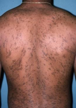
Figure 1. Erythema dyschromicum perstans (EDP). EDP presents with widespread well-demarcated and slowly expanding blue-gray macules. The lesions rarely cause mild itch and demonstrate an erythematous border in the acute inflammatory phase. While involvement of the face, neck, limbs, and trunk has been described most commonly, a unilateral presentation has also been reported.
The histological features of EDP depend on the age of the lesion biopsied. Early on, papillary dermal edema and basal layer vacuolation with a perivascular infiltrate is noted. Later, the inflammatory cell infiltrate becomes sparse, but melanin incontinence and melanophages are noted in large amounts in the dermis, particularly the deep dermis.
Unfortunately, as the name suggests, the condition is chronic and a challenge to treat. Unsurprisingly, while hydroquinone and tretinoin are helpful in pigmentary conditions, such as melasma and post-inflammatory hyperpigmentation, they have not demonstrated significant lightening in EDP, likely due to the dermal location of abnormal pigment. Dapsone has been reported to lighten pigmentation and even completely resolved it when taken for 8-12 weeks at a dose of 100 mg daily. While clofazamine demonstrated improvement in a small study (100 mg daily), the anecdotal experience among pigmentary disorders experts globally has not been so positive. Unfortunately, while many other treatments have been evaluated, including topical steroids and chloroquine, none have resulted in appreciable lightening. Most recently, laser surgery has been studied by a group in the Netherlands, who concluded fractionated laser was unsuccessful for treatment of EDP. Examination of the literature highlights the need to more clearly define this entity and to thoroughly evaluate the efficacy of treatment options.
Lichen planus pigmentosus (LPP)
LPP is a rare variant of lichen planus usually seen in middle-aged individuals with more deeply pigmented skin (e.g. South Asians, South-East Asians and the Arabic population). Though this is most commonly seen in photo-exposed sites, such as the head (forehead and temples) and neck, the symmetrical brown to gray-brown macules and patches are often seen in skin folds such as the axillae (LPP-inversus) and occasionally the mucosa. Unlike EDP, LPP lacks clinical signs of inflammation and does not have lesions with erythematous borders.
The exact cause of LPP is unknown, although ultraviolet light has been implicated due to the location of the lesions. Mustard and amla oils have been reported as possible causes in some retrospective studies. These oils are used routinely in India in hair care preparations, massage oils and for cooking. Allyl thiocyanate is a potential photosensitizer in mustard oil and may be a pathogenic agent in LPP.
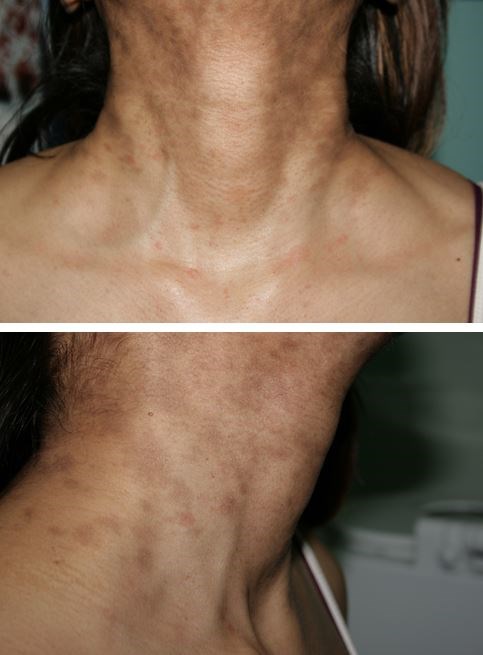
Figure 2. Lichen planus pigmentosus (LPP). Histopathological evaluation of lesional skin reveals epidermal atrophy and basal layer vacuolation with a perivascular lymphocytic infiltrate. The dermal melanophages are situated in the superficial dermis in LPP in contrast to EDP in which they are in the deep dermis.
LPP undoubtedly presents a therapeutic challenge. While some cases gradually resolve over many months, many persist for years or even decades. Sunscreen and sun-protection are key components in the treatment of LPP. Various small studies reveal topical tacrolimus (0.03% twice per day), topical and systemic corticosteroids and topical vitamin A may be helpful in LPP. Combinations of the above-mentioned therapies have also been studied to prevent progression of LPP, but advances clearly need to be made in the LPP therapeutics arena.
The blurred lines between LPP and EDP clearly demonstrate the need for a large scale, multi-center study to further delineate these two conditions etiologically, morphologically and histologically. A summary of the treatment options currently used in the management of EDP and LPP are shown below in Table 2. However, large, double-blinded randomized studies are needed to more accurately assess the success of therapeutic interventions.
Table 2. Summary of therapeutic options for EDP and LPP.
EDP | LPP |
Photoprotection | Photoprotection + avoid potential triggers |
Potent topical corticosteroids | Potent topical +/- oral corticosteroids - especially if disease is actively expanding, itching |
Hydroquinone 4% or more | Tacrolimus 0.03% |
Tretinoin | Tretinoin |
Dapsone | Hydroquinone 4% or more |