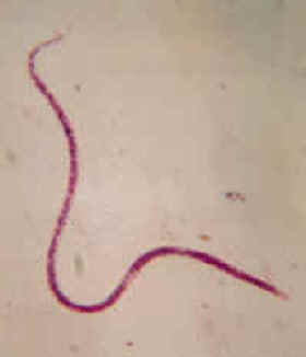
Filaria
Filaria medinensis
patient (carrier) but rarely observed in
clinical cases making it difficu
lt for precise
diagnosis & management
Filaria is a parasitic worm from the Nematoda class. Its length can vary from 2 mm to 1.3 m. It lives in the tropics and subtropics. The adult individuals parasite in different parts of the human body (where they get through contaminated water or food) but usually prefer the limbs. They live under the skin where they make protuberances. This leads to swellings and inflammations, later boils. If the worm tears while pulling out of the skin, occur severe inflammation and a poisoning, sometimes with a lethal end. These symptoms are obviously due to the body fluids of the worm because if it is pulled out the whole, the recovery is very fast.
In Guatemala and Mexico there is a similar parasite which can reach a length up to 50 cm. Its poison causes visual impairments, even loss of sight, convulsions, muscle pain, sleeplessness. After removing the worm, the recovery is almost immediate.
FILARIASIS
A major public health problem Human lymphatic filariasis is caused by the infection with major nematode parasites Wuchereria bancrofti and Brugia malayi. The disease is quite prevalent in the developing tropical countries. About 120 million people are infected all over the world, of which 43 million are affected with overt physical disabilities from filarial infection.
In India around 412 million people are living in bancroftian endemic areas with 31 million people are estimated to be harbouring microfilariae and about 20 million people suffer from clinical manifestations of the disease, with about 7.5 million of lymphoedema cases and 13 million of hydrocele cases (1). Further, millions suffer with occult filarial infection in endemic areas without diagnosis. Filariasis is prevalent in all states and union teritories except Jammu & Kashmir, Himachal pradesh, Punjab, Haryana,Rajasthan,Meghalaya,Mizoram, Nagaland, Manipur, Tripura & Sikkim.
Although the disease is never directly fatal, it is a debilitating one responsible for considerable morbidity and social stigma. In 1995, the World Health Organization (WHO) identified filariasis as world’s second leading cause of permanent and long term disability next only to mood affecting disorders (2). The global burden of lymphatic filariasis was estimated to be at least 850,000 DALYS (Disability Adjusted Life Years Lost) and India contributes 38% of the global disease burden. In endemic areas out of 1500 million children in the age group of 0-14 years,14 million harbour microfilaraemia, while 1 million suffer with lymphoedema and 2 million with hydrocele.
Wide spectrum of clinical manifestations in filariasis which need diagnosis and OpDEC therapy*
Acute & Chronic
- Fever with chills and rigors
- Lymphoedema with pain
- Lymphadenopathy (Cervical, Axillary, Inguinal& Generalised)
- Chyluria / Haematuria
- Funiculitis
- Epididymoorchitis
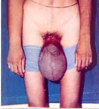
- Hydrocele
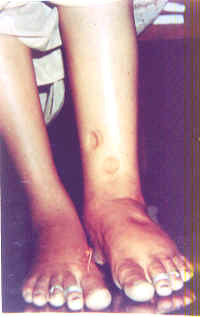
- Elephantiasis
Occult
- Pulmonary eosinophilia
- Mono & Polyarthritis
- Tenosynovitis
- Glomerulo nephropathy
- Retroperitoneal lymphangitis (Acute abdomen)
- Central serous retinopathy
- Iridocyclitis, recurrent scleritis & macular oedema
- Endomyocardial fibrosis
- Urticaria
- Recurrent URI
- Asthmatic bronchitis

Optimal DEC therapy (6mg / kg body wt / day for 21 days each month for 3-12 months). The period of DEC treatment is determined based on immunomonitoring of infection.
Filariasis is a mosquito borne disease. The microfilariae sucked up by mosquito from infected man develop to infective larvae in 1014 days. Mosquito harbouring infective stage larvae bite and infect a fresh host (man).
Larvae migrate into Lymphatics,where they settle,grow & mature into adult worms.Followed by fertilization the gravid female delivers microfilariae into blood circulation and thus setting stage ready for fresh transmission to more and more people.
Clinical Manifestations
Lymphatic filariasis is characterised by a wide spectrum of clinical manifestations
Asymptomatic microfilaraemia
This stage is characterised by the presence of microfilariae in peripheral blood during night but without any overt clinical manifestations of filariasis. It is possible that some of these have hidden lymphatic pathology in the form of dilatation at high risk of developing disease.
Acute manifestations
These are characterised by recurrent attacks of fever associated with inflammation of the lymphnodes and lymphvessels - acute adenolymphangitis (ADL). Various factors related to parasite like release of toxins, immune response in infected human host , trauma in the affected area and secondary bacterial and / or fungal infections play a role in causing the ADL attacks. The inflammed lymphatics of the male genitalia lead to funiculitis or epididymoorchitis.
Chronic manifestations
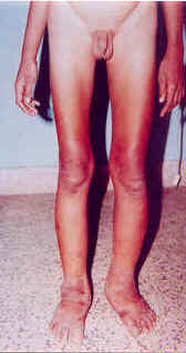
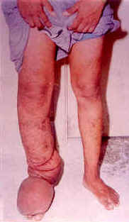
Major chronic clinical manifestations are hydrocele, lymphoedema and elephantiasis.
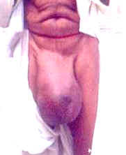
While hydrocele is the commonest genital manifestation in the male population, chronic epididymitis, funiculitis and lymphomatous thickening of scrotal skin can be the other genital manifestations.Lymphoedema commonly affects the lower limbs, some times the hands, and rarely the genitals and breast in females . There may be repeated attacks of ADL.
Lymphoedema progresses from pitting oedema (reversible on elevation) and then non-pitting oedema followed by increase in oedema fluid volume, resulting in fibrous tissue formation, followed by skin thickening, ulceration, nodule formation and disfiguration (elephantiasis).
Chyluria is prevalent in some endemic areas.
Occult manifestations
Some filarial patients do not have any classical clinical manifestations and do not show microfilariae in peripheral blood, although the parasites may occur in internal organs or tissues.
The major occult filarial manifestations include tropical pulmonary eosinophilia (resulting from allergic reaction to parasite and characterised by nocturnal paroxysmal cough and hypereosinophilia), glomerulopathies, endomyocardial fibrosis, monoarthritis, tenosynovitis, acute abdomen, central serous retinopathy (CSR), iridocyclitis, urticaria, haematuria etc. in adults.
Pulmonary eosinophilia associated with cough, asthmatic bronchitis, arthritis, recurrent URI, pneumonitis, etc. were observed in children in filaria endemic areas responding to DEC therapy.
Diagnostics
The presice diagnosis of filariasis is presently based on the demonstration of the parasites in the night blood is incovenient, inconsistent and not sensitive in detecting low microfilaraemia. Further, microfilariae are not normally seen in peripheral blood in acute and chronic infections as well as occult filarial infections, making it difficult to diagnose clinically for successful treatment. Most of the clinicians rely on their clinical acumen in the diagnosis of clinical filariasis.
Immunodiagnosis:
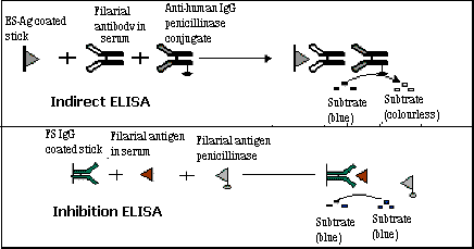
SEVA FILACHEK (IgG & Ag assay) dipstick based ELISA system has been explored in several ways at MGIMS using penicillinase enzyme, microfilarial antigen and filarial antibodies in diagnosis of filarial infection in different clinical groups(3). The detection of IgG antibody (titre 1:300 & above) against specific microfilarial antigen was found to be useful for detecting microfilaramic and most of the clinical filarial cases. Free and immune complexed filarial antigen (titre 1:300 & above) are detected using filarial serum immunoglobulin G (FSIgG). Filarial antigen detection was found to be more useful in epididymoorchitis and allergic state such as Tropical eosinophilia . The test system showed a sensitivity and specificity of about 80% (4,5).
The antibody detection test is based on the detection of specific IgG antibody to Brugia malayi mf ES antigen by indirect penicillinase ELISA. The mf ES antigen coated CAM sticks are incubated with diluted (1:300) filarial sera followed by antihuman IgG penicillinase conjugate. After washing, when the sticks are finally incubated with blue coloured starch-iodinepenicillin ‘V‘ substrate, the disappearance of the colour earlier than control indicates the presence of filarial antibody. The free and IC antigen detection is done by inhibition ELISA.The CAM sticks, sensitized with FSIgG isolated from clinical patient’s sera are incubated with appropriate test sera (1:300) followed by mf ES antigen penicillinase conjugate. The presence of filarial antigen is indicated by the persistence of the blue colour of starch-iodine-penicillin ‘V‘ substrate. The free antigen detection assay was found to be useful to detect microfilaraemic patients (80%), whereas, IC antigen detection was useful to detect clinical filariasis viz; chronic filariasis (80%), acute filariasis (88%) & occult filariasis (78%).
References:
- National Institute of Communicable Diseases – revised strategy for control of lymphatic filariasis in India (Recommendations of the WHO sponsored Workshop) (1996) ; New Delhi, 4-5 January.
- The World Health Report – Bridging the gap. Report of the Director General, WHO (1995); Geneva – 3.
- Harinath, B.C ., Malhotra, A., Ghirnikar, S. N., Annadate, S.D., Isaacs, V.P. & Bharati , M.S. Field evaluation of an enzyme linked immunosorbent assay using Wuchereria bancroti mf ES antigen for bancroftian filariasis ., Bulletin of World Health Organisation (1984) ; 62 , 941-944.
- Alikhan , A., Parkhe, K. A., Reddy M.V.R. & Harinath , B.C., Filarial antigen, antibody and circulating immune compexed antigen levels in bancroftian filariasis by stick ELISA . The National Medical Journal of India (1990); 3 (6) 265-268.
- Alikhan , A ., Padigel , U . M., Ramarao, B. V., Adwani, B., Reddy M.V.R., Chaturvedi, P. & Harinath, B.C., Filarial antibody and antigen detection in different clinical conditions in an endemic area , IJCB.; (1994); 9 : 1;31-34.
- Harinath , B.C., Reddy, M.V.R., Padigel , U. M., Devi, K.K., Alli, R. & Mehata, V. K ., Seva-Filacheck for immunomonitoring of clinical and occult filarial infections , J.of MGIMS ; Sept. (1996); 52-56.
- Harinath, B.C., Reddy, M.V.R., Bhunia, B., Bhandari, Y.P., Mehta, V.K., Chaturvedi,P., Prajapati, N.C. & Gupta, R.K.C. Filaria associated clinical manifestations in children in an endemic area and morbidity control by immunomonitoring and optimal DEC therapy: Sevagram experience, IJCB; (2000),15 (suppl.); 118-126.
- Harinath, B. C. & Reddy , M.V.R. ,Diagnosis and immunomonitoring in the successful management of bancroftian filariasis , J. of Parasitic Diseases; (1997), 21 ; 41-51.
- Alli,R.,Kulkarni,S.,Reddy,M.V.R.& Harinath, B.C. Evaluation of SEVA FILACHEK immunoassays and rapid ICT – filariasis test for detection of bancroftian filariasis, IJCB; (2001), 16(2),207-210.
- Current Medical Diagnosis and Treatment 2001, 1469.
- ICMR Bulletin: Vol. 28: No. 5, May, 1998.
- Management of lymphatic filariasis - A manual for clinicians. Vector Control Research Centre, Misc. Publ. (21) 1997.